
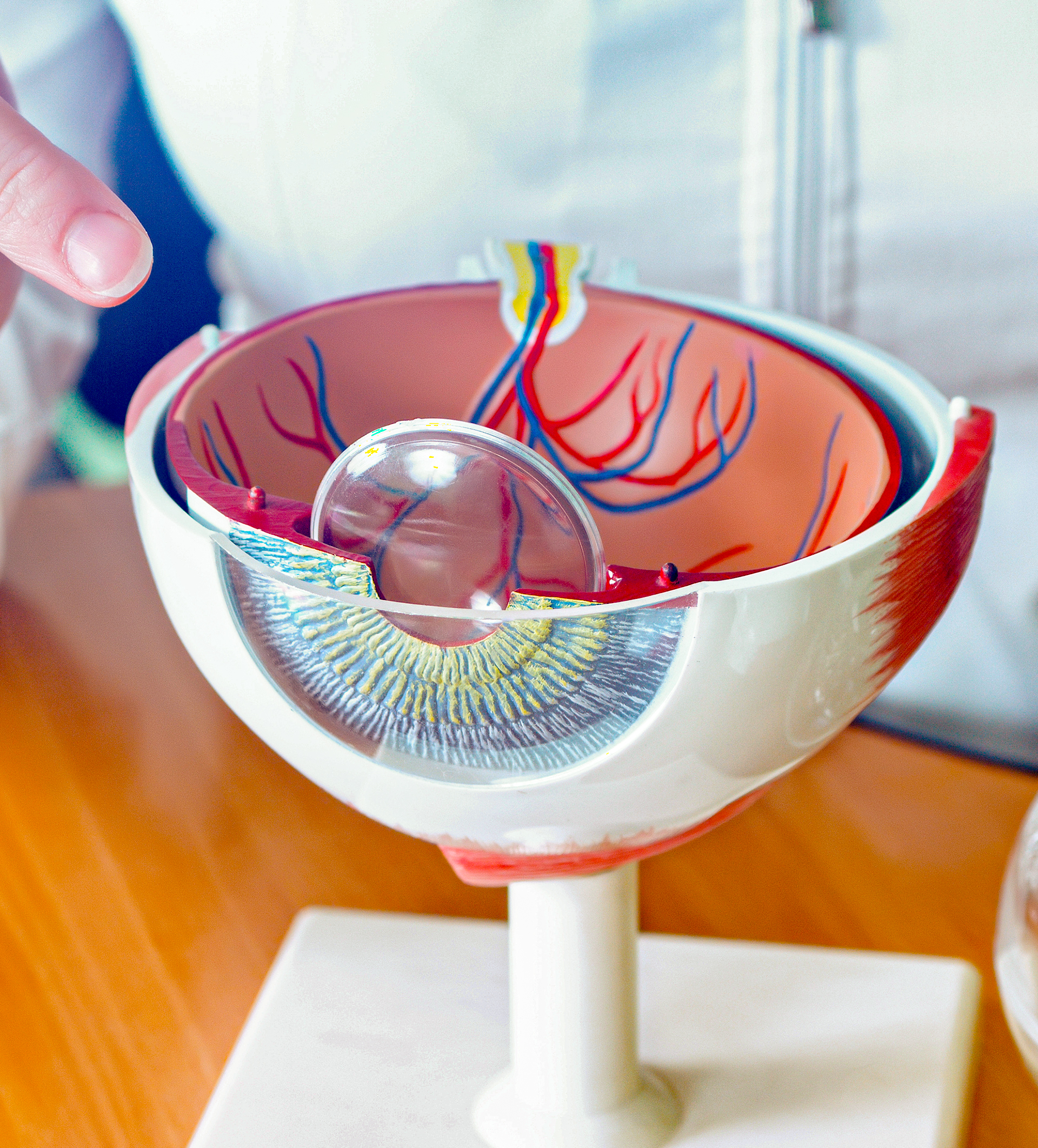
Retinal detachment is a serious eye condition that can lead to permanent vision loss if not treated promptly. It occurs when the retina, the thin layer of tissue at the back of the eye, separates from its supportive tissue. While retinal detachment can happen suddenly and without warning, understanding the causes and taking preventive measures can reduce your risk. Additionally, early diagnosis and the latest treatment options can help preserve vision. In this blog, we’ll explore retinal detachment's causes, prevention, and management.
Causes of Retinal Detachment
There are three main types of retinal detachment: rhegmatogenous, tractional, and exudative. Each type is caused by different underlying factors:
Rhegmatogenous Retinal Detachment: This is the most common form of retinal detachment. It occurs when a tear or hole forms in the retina, allowing fluid to seep underneath and cause separation. Factors that can lead to this type of detachment include:
- Aging can cause the vitreous gel inside the eye to shrink and pull on the retina.
- Severe myopia (nearsightedness) increases the risk of retinal thinning or tearing.
- Previous eye surgery, such as cataract removal, which can lead to complications involving the retina.
- Eye injuries, which may result in retinal tears or detachments.
Tractional Retinal Detachment: This type occurs when scar tissue on the retina contracts, pulling the retina away from the back of the eye. It is commonly associated with conditions like diabetic retinopathy, where uncontrolled blood sugar levels lead to the growth of abnormal blood vessels and scar tissue.
Exudative Retinal Detachment: This form occurs without any tears or holes in the retina. Instead, fluid builds up beneath the retina due to inflammation, tumors, or other medical conditions such as central serous retinopathy.
Preventive Measures
While not all retinal detachments can be prevented, there are steps you can take to reduce your risk, especially if you are in a high-risk group (e.g., older adults, individuals with myopia, or those with a family history of retinal problems):
- Regular Eye Exams: Early detection is crucial in preventing serious complications. If you have a history of retinal issues or are at higher risk for retinal detachment, ensure you have regular comprehensive eye exams with a retinal evaluation.
- Protect Your Eyes: If you engage in high-risk activities, such as contact sports or work in environments with a high risk of eye injury, wear protective eyewear to prevent trauma.
- Manage Health Conditions: Keep conditions like diabetes under control to avoid complications like diabetic retinopathy, which can increase the risk of retinal detachment. Proper management of blood pressure and cholesterol can also contribute to better eye health.
- Watch for Symptoms: Warning signs of retinal detachment may include flashes of light, a sudden increase in floaters, or a shadow or curtain-like appearance across your field of vision. If you experience any of these symptoms, seek immediate medical attention.
Treatment Options for Retinal Detachment
The key to treating retinal detachment is prompt intervention. Several advanced surgical techniques are available to reattach the retina and restore vision. The appropriate treatment depends on the severity and type of detachment.
Laser Surgery or Cryotherapy: For small tears or holes, laser photocoagulation or cryotherapy (freezing) can be used to seal the retina and prevent detachment. These procedures are minimally invasive and often performed in an outpatient setting.
Pneumatic Retinopexy: This is a procedure where a gas bubble is injected into the eye to push the detached retina back into place. The patient must maintain a specific head position to keep the bubble in place until the retina reattaches.
Scleral Buckling: This procedure involves placing a silicone band around the eye to push the wall of the eye inward, helping the retina reattach. Scleral buckling is a more invasive option but highly effective for larger or more severe detachments.
Vitrectomy: In more complex cases, a vitrectomy may be performed to remove the vitreous gel and replace it with a saline or gas bubble to hold the retina in place. This option is often used in cases involving tractional or severe rhegmatogenous detachments.
Retinal detachment is a serious condition that requires immediate medical attention to prevent permanent vision loss. Understanding the causes, taking preventive measures, and seeking timely treatment can help preserve your vision. At Coastal Eye, we specialize in diagnosing and treating retinal conditions with the latest techniques. If you are experiencing symptoms of retinal detachment, contact us right away for a consultation.